Each 3C Patch® Kit is packed in an individual box and contains all the components needed to produce a 3C Patch®.
The only missing ingredient is an 18 ml sample of the patient’s own blood which is taken at the point of care.
The 3C Patch® Kit is a procedure pack providing the single-use components needed for the production of the 3C Patch®
Return
< Home

Make A Patch

At Reapplix, we have established that one of the keys to successful wound treatment lies in the body’s capacity to heal – and each patient is central to that process. Using just a small sample of the patient’s blood – nothing else

100% Autologous - no additives or reagents

The 3C Patch® Kit is a procedure pack providing the single-use components needed for the production of the 3C Patch®
Each 3C Patch® Kit is packed in an individual box and contains all the components needed to produce a 3C Patch®.
CENTRIFUGATION
The proprietary high speed centrifugation step separates cells and is a unique part of the patented 3C Patch® Process
COAGULATION
Fibrin polymerizes during the coagulation step monitored by the centrifuge’s built-in optical sensor
COMPACTION
Compaction forms the robust 3C Patch®
The unique process provides autologous wound management at the point of care, meaning a 100% unique patch prepared from the patient’s own blood. This fully automated process takes approximately 20 minutes:

Patented fully automated centrifugation process

15-20 minutes is

the approximate time it takes

to produce

a 3C Patch®

18ml of blood is collected into the 3C Patch® device by vacuum. The device is placed into the centrifuge to generate a 3C Patch®.
The blood cells are separated into three layers using a proprietary process controlled by the centrifuge.
The important components of blood reaching a wound are; immune cells, platelets and growth factors. These components help orchestrate the wound healing response.
The filter is released to compact the leukocytes, platelets and fibrin creating the 3-layer patch.

Transfer the 3C Patch® to the patient

Histological stain highlighting the three layers of the 3C Patch®

Histological image highlighting the three layers of the 3C Patch.

More than 800 different growth factors, cytokines and proteins have been detected in the 3C Patch® by multiplex analysis

Open the device and remove the 3C Patch®.

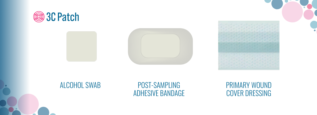
Place the 3C Patch® filter side up on a sterile absorbing surface.

Apply the filter side (checkered side) of the patch directly to the wound surface. The 3C Patch® may overlap intact skin.

Cover the 3C Patch® with the supplied non-adherent primary wound cover dressing. Apply a secondary absorbent dressing (free choice, depending on the amount of exudate).

Back to Top

Clinical Evidence

One of few evidence-based wound management procedures

The 3C Patch® is clinically proven to significantly increase the chance of healing of chronic wounds associated with diabetes.

A promise backed with evidence. Proven results including a large RCT published in The Lancet Diabetes & Endocrinology1 and supported with additional solid scientific and clinical data.

Randomized controlled (n= 269) trial published in the Lancet Diabetes & Endocrinology, 2018.

One of few evidence-based wound treatments, the 3C Patch® is clinically proven to significantly accelerate wound healing of hard-to-heal diabetic foot ulcers.

3C Patch is recommended as adjunctive treatment in hard to heal diabetic foot ulcers.

Wound types: Wagner 1,2,3 ABI down to .5
1st Patch Application: Clean and debride the patient's 
wound to allow the cells
 and signaling molecules in 
3C Patch to interact with 
living tissue.
2nd and 3rd application: After the 2-3rd application
an increase in granulation
tissue is usually seen.

Request a free copy of the 3C Patch Clinical Trial Overview.

3C Patch Study Design3C Patch Intervention3C Patch Patients

Scan code with your own device:

Back to Top
Patch Process screensaverWound Healing
screensaver